Macular Degeneration

Macular Degeneration: A Tough Nut to Crack. Macular degeneration is the most common cause of legal blindness (less than 20/200 in both eyes) among people over age 60 in the United States, afflicting millions.
Understanding the Macula
The macula is located in the middle of the retina. The center of the macula is the fovea, which controls central vision, the sharpest portion of the entire visual field.
We use the macula and fovea for reading, cooking, watching TV, playing tennis, distinguishing colors, and just about everything that requires perception of fine details.
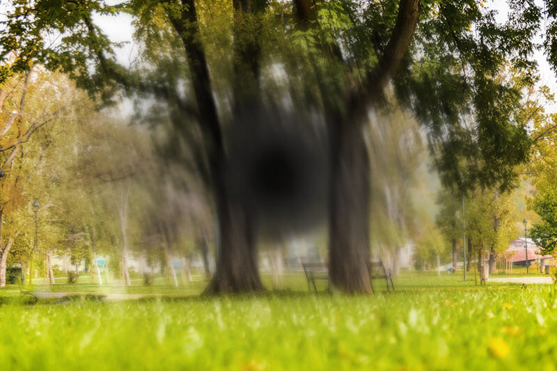
Macular degeneration destroys the most important part of the retina. Macular degeneration generally does not affect peripheral vision, so patients do not go completely blind.
Macular degeneration is a diverse group of diseases, the most common of which is age-related macular degeneration (ARMD or AMD).

What Causes Macular Degeneration?
AMD is classified into the "dry" and "wet" forms. The much more common dry form is characterized by degeneration of the macula and tissues that support the function of the macula.
Dry macular degeneration does not respond to current available treatments.
Fortunately, not everyone will suffer from significant vision loss.
Occasionally, dry macular degeneration transforms into the more serious wet form.
Vision may deteriorate rapidly and suddenly from wet AMD.
Abnormal blood vessels and membranes proliferate under the macula, causing bleeding, swelling, and eventually scarring.
Depending on the location of the macular degeneration (whether it is directly under the very center of the macula) and the stage of the disease, different treatments may improve or stabilize vision.
How the disease actually occurs remains a mystery.
Risk Factors
- Age. The risk of AMD rises steadily with age.
- Caucasian race, especially those with blue eyes.
- Smoking. Smokers have much higher risk of developing AMD and suffering vision loss from AMD.
- Diet high in fat and cholesterol.
- High blood pressure.
- Genetics. The genetics appears very complicated. Although the risk of blood relatives of an AMD patient is higher than that of the general public, the risk cannot be easily quantified.
Symptoms
- Central vision loss
- Distorted vision — straight lines appear wavy
- Shady or blurry areas near the center of vision
- Sudden onset of symptoms is possible
- Often affects one eye initially
- Mild cases may be asymptomatic
How Do You Diagnose Macular Degeneration?
Macular degeneration is diagnosed by examining the retina. The patient may be completely asymptomatic at the earlier stages of the disease. Tests such as optical coherence tomography (OCT), which uses light to scan the macula to obtain a high definition, cross-sectional picture, and fundus photography are done non-invasively to document and follow the disease. Fluorescein angiography, which requires injection of a dye into a vein in the arm, is used to identify the exact location of wet macular degeneration to pinpoint the best treatment.
How Does Macular Degeneration Get Treated?
There is no effective conventional remedy for dry AMD. Wet AMD may be treated with laser treatment (in cases where the wet AMD is not directly under the center of the macula) or injection of medication into the eye, such as Avastin, Lucentis, and Eylea. While not without discomfort, the injections are well-tolerated by most patients. However, they do not benefit every patient. Treatment is often highly individualized, so discuss with your eye doctor about your specific situation. Photodynamic therapy represents an uncommonly used alternative.
Prevention and Stabilizing Macular Degeneration
- Quitting smoking.
- Controlling high blood pressure by exercise, diet, and medications.
- Avoiding excessive ultraviolet light (stay out of the sun or wear glasses with UV light protective coating).
- Consuming a healthy diet rich in fruits, vegetables, and dietary fiber. Restriction of animal fat and cholesterol may be helpful.
- Taking lutein, zeaxanthine, and/or the AREDS (Age-Related Eye Disease Study) formulas (AREDS I and AREDS II). Seek advice of your eye doctor and medical doctor before initiating these supplements, especially if you smoke or have a history of smoking.
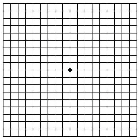
Once you have been diagnosed with macular degeneration, you should perform periodic Amsler grid exam:
- Hold the grid 14 inches away; put on reading glasses.
- Cover one eye.
- Focus on the black dot in the center of the grid.
- If any areas on the grid are hazy, black, distorted, or discolored, or if any lines are crooked, or if the exam appears to have changed since the last time, notify your doctor.
- Repeat with the other eye.
Have questions about macular degeneration?
Our experienced team is ready to help you see clearly.
Schedule a Consultation